When we want to examine the brain of a person noninvasively by Computed Tomography (CT) or MRI, we get a ‘snapshot’ of the anatomy (or pathology, if any) of the subject’s brain. We are however clueless as to its functional aspect. fMRI or Functional Magnetic Resonant Imaging allows us to do just that. The difference is not unlike a ‘still picture’ versus a ‘video of a moving train’. PET scans, previously described, also can asses the functional state of the brain.
Whenever we do a task, think, dream, memorize, speak or see things, the brain is not activated as a whole; but only certain portions of it are activated. Activation, here, means increased metabolic activity of neurons in certain areas of the brain. Naturally, these ‘metabolically active’ neurons would demand more energy which would power them. The blood supply to these areas increases as a result of this metabolically driven vasodilation. The arteries then bring in glucose and oxygen with them, with Oxygen being transported in the form of Oxyhemoglobin (oxygenated hemoglobin or HbO2). Neurons on the other hand use up the oxygen contained in the blood, thereby reducing it to de-oxyhemoglobin orsimply Hb. However, the alteration in tissue perfusion exceeds the extraction of oxygen by the neurons, so the concentration of deoxyhemoglobin within ‘the areas’ decreases. This causes molecular inhomogeneities in the magnetic field.
Oxyhemoglobin is diamagnetic, meaning that they align perpendicularly to magnetic field lines. On the other hand, deoxyhemoglobin is paramagnetic, i.e. it aligns parallely and proportinately with the intensity of the magnetic field. This causes the inhomogeneity within the magnetic field (magnetic susceptibility) in the tissue sampled. This inhomogeneity is exploited in fMRI in terms of decay of transverse magnetization, T2*, with longer T2* values in HbO2 blood and shorter values in Hb (paramagnetic) blood.Since this stems from the oxygen content in blood, fMRI is also known as the BOLD ((blood oxygenation level dependent) effect.
The machine is essentially the same as the MRI machine with echo planar imaging technology that permits faster imaging due to faster gradient switching, improved algorithm and faster CPU processing power. The patient/subject is placed inside the magnetic chamber and MRI signals are acquired, Fourier transformed and corrected for artifacts. Finally the computer reconstructs a 3D fMRI image out of this.
As is obvious, we can learn about the motor areas of a patient by asking him to grasp an object or giving him any motor task and noticing which area(s) of the brain lights up. A neurosurgeon can then be cautious about not hurting these areas. Similarly, the mapping will help spare motor and other vital areas like auditory, visual and language areas from damage in radiotherapy procedures, in addition to neurosurgery. It can also detect occult Alzheimer’s disease and cognitive deficits including those of the autism spectrum anddyslexia (reading disorder).
fMRI can also be employed to ‘read peoples’ minds’, thoughts, intentions including lie detection. Watch the video below which explains how an FMRI scan is done and interpreted.
Thus the legal and forensic implications are obvious. However, in fMRI, correlation doesn't always mean causation. Whatever it may be, it seems that fMRI is very much here to stay, both in the clinics as well as in cognitive neuroscience research. It may also be combined with tractography, MRI or other diagnostic radiologic modalities.
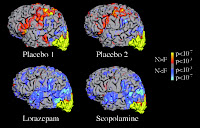
Hardenbergh et al combined Tractography techniques with fMRI, using a technique capable of rendering multiple color-coded functional activation volumes and fiber tract bundles. Many pharmacologically active drugs have effect on memory impairment, which can be seen in ‘telltale’ fMRI scans. Sperling et al studied the effects of lorazepam (a benzodiazepine) and scopolamine (an anticholinergic drug once used as ‘truth serum’ by the CIA)
 on healthy volunteers and found that theydid impair memory and their functional coordinates could be reproducively mapped on fMRI scans (see figure on the left). I still shudder at the thought of what happened during my PG exam when I took a benzodiazepine.
0 comments:
Post a Comment